2026-11-03
Retinal Detachment🚑 Emergency Surgery❗ When Vision Looks Like a Black Curtain Has Been Drawn❕ or Black Spots Float Before Your Eyes📛
Explains retinal detachment causes and symptoms such as a black curtain over vision or floaters, surgical options like scleral buckling and vitrectomy, and why early diagnosis matters.
Retinal Detachment
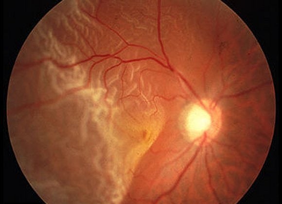
Fundus photo of retinal detachment: the pale, wrinkled area on the left is the detached retina.
This means the retina has come loose.

Fundus photo of an extensive retinal detachment, with the retina lifted in folds around the optic disc.
In cases of partial retinal detachment, laser treatment alone may sometimes be enough, but most cases require emergency surgery.
I performed this surgery countless times during my time as a clinical fellow at a university hospital, roughly one case every two days.
Although retinal detachment can be subdivided into several types in academic terms,
it usually refers to rhegmatogenous retinal detachment (RRD), which is caused by holes or tears in the retina.
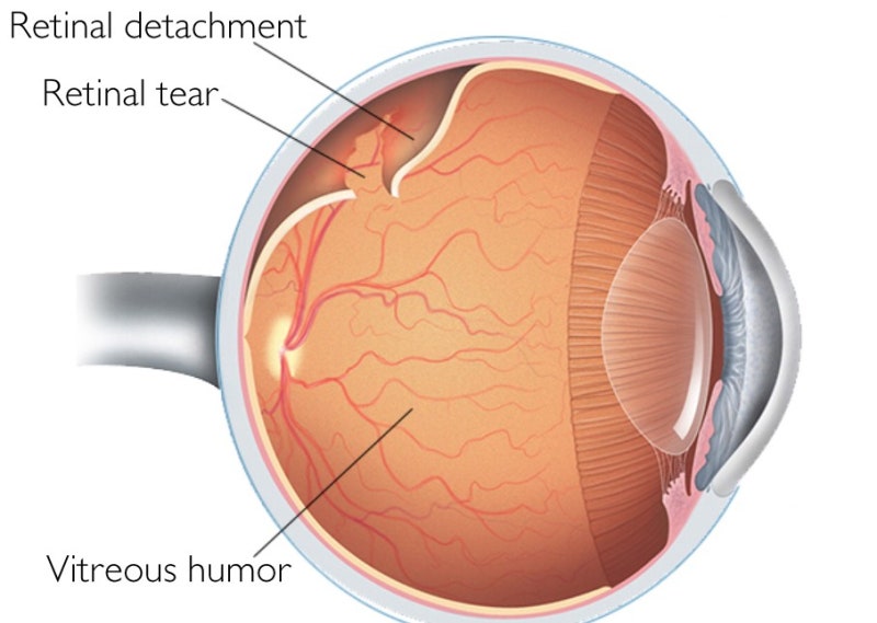
Eye cross-section diagram labeling a retinal tear, the retinal detachment and the vitreous humor.
The causes of holes or tears in the retina are as follows:
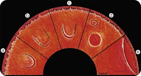
Illustration of different types of retinal tears and holes, labeled a to e.
In highly nearsighted eyes, the peripheral retina may naturally thin and develop holes,
and tears can also occur when the vitreous separates from the retina due to posterior vitreous detachment (PVD), pulling on the retina and tearing it.
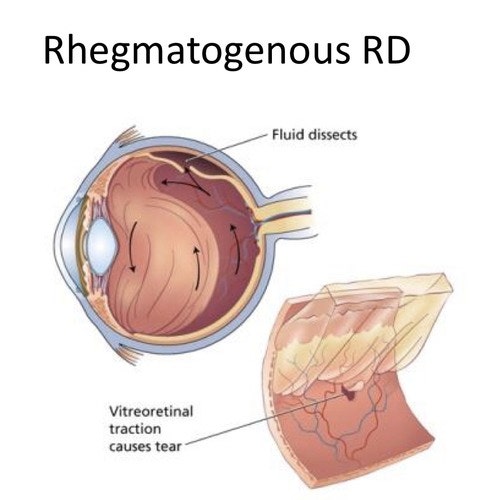
Diagram of rhegmatogenous retinal detachment: vitreoretinal traction causes a tear and fluid spreads under the retina.
The retina is attached to the underlying choroid and must adhere firmly to the sclera, the wall of the eyeball, like wallpaper.
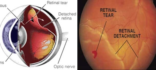
An eye diagram and a fundus photo, each marking a retinal tear and the detached retina.
The mechanism of retinal detachment is this: the retina develops a hole or tear, fluid seeps through the gap, and the retina separates from the wall.
The retina is a thin, wallpaper-like sheet of optic nerve fiber bundles connected to the brain,
and when it detaches, visual signals can no longer be transmitted, so the detached area appears black.
Because it typically progresses from the periphery,

A street scene partly covered by a dark, curtain-like shadow, illustrating vision loss from retinal detachment.
Patients often describe it as “a black curtain covering their vision from the edges.”
If retinal detachment is caught early and only a partial detachment exists around the tear,
a barrier laser procedure, literally creating a barrier with laser photocoagulation to keep the detachment from progressing, may be attempted.
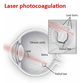
Laser photocoagulation diagram: a laser beam enters the eye and laser burns are placed around a retinal tear.
To put it simply, it's like stapling the spot where the wallpaper has torn and started to peel, to hold it in place.
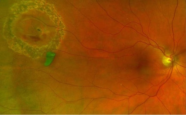
Fundus photo showing a ring of laser marks surrounding a lesion on the retina.
Unfortunately, such good fortune is rare,
and most cases require surgical treatment.
The types of surgery performed for retinal detachment include:
One is scleral buckling/encircling, in which a silicone band is placed on the outside of the eye to press on the hole, combined with cryopexy (freezing coagulation).
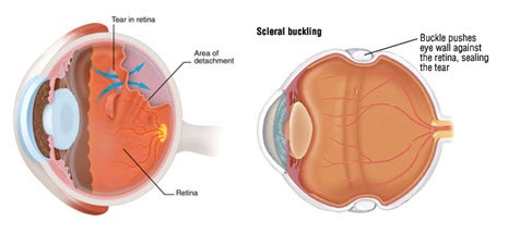
Left: a retinal tear with detachment. Right: scleral buckling, where a buckle pushes the eye wall against the retina to seal the tear.
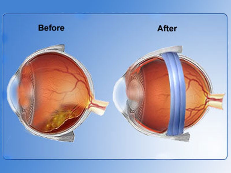
Before-and-after illustration: a detached retina, then the eye with a scleral buckle band placed around it.
There is also pars plana vitrectomy (PPV), in which surgical instruments are inserted into the eye to directly drain subretinal fluid, followed by laser photocoagulation and gas or silicone oil injection.
For giant tears or severe retinal detachment, both of these surgeries may be performed together.
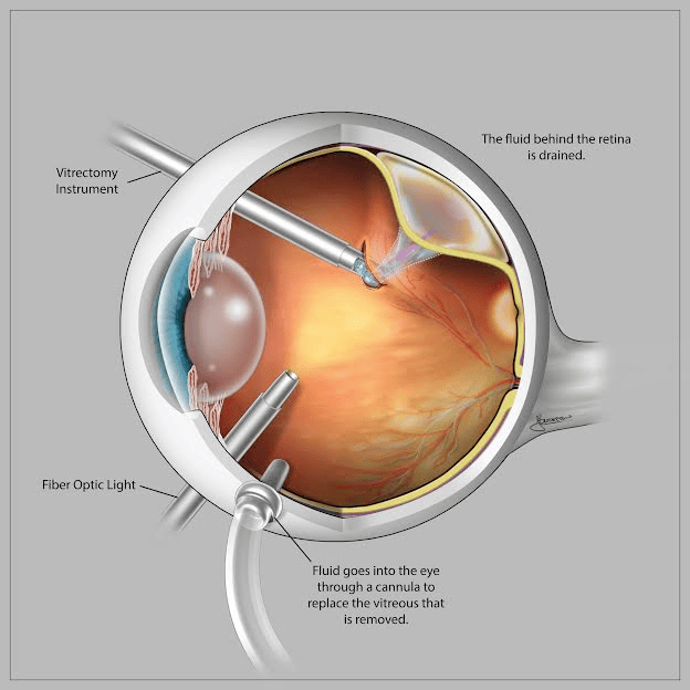
Vitrectomy diagram: instruments enter the eye, fluid behind the retina is drained, and fluid replaces the removed vitreous.
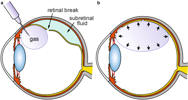
Gas injection diagram: (a) gas is injected near the retinal break; (b) the expanded bubble presses the retina back into place.
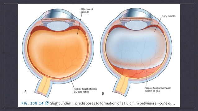
Because gas or silicone oil floats upward in the fluid inside the eye due to buoyancy, this buoyant force can be used to apply pressure to the desired area of the retina.
Based on this principle, retinal detachment is treated surgically.
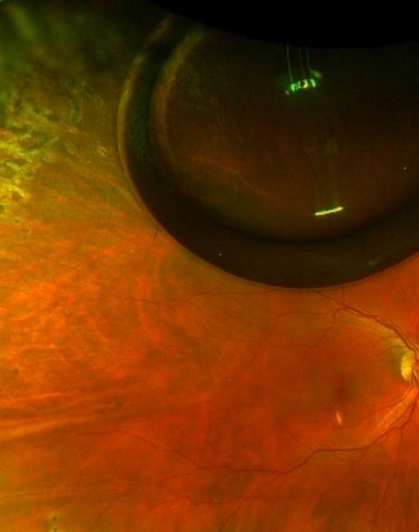
Wide-field fundus photo; the dark round shape at the top is a gas bubble inside the eye.
Diagram comparing an eye filled with silicone oil (A) and one with a C3F8 gas bubble (B), each with a thin film of fluid.
As such, accurate diagnosis and appropriate treatment at an early stage are essential for retinal detachment.
Even after successful surgery, retinal detachment can recur due to trauma or other factors, so patients should take special care.
In particular, if you notice floaters (myodesopsia), dark spots drifting in front of your eyes, they may be related to a retinal hole or tear, so be sure to get a detailed retinal exam at an eye clinic!
For more details on this topic, please refer to the Seongmo Jin Eye Clinic website.
If you are curious about your eye condition or experiencing discomfort, you can consult Park Jin-hyung, Director of Seongmo Jin Eye Clinic, who serves as a retina clinical fellow and outpatient professor at Seoul St. Mary's Hospital. ^^
#SeongmoJinEye #JinEyeClinic #Ophthalmology #Presbyopia #Cataract #Multifocal #Retina #Glaucoma #DryEye #DryEyeSyndrome #DryEyes #MacularDegeneration #RetinalDisease #CataractSurgery #GangnamEyeClinic #SeongmoEyeClinic #PresbyopiaSurgery #OphthalmologyProfessor #RealEyeClinic #LASIK #LASEK #GangnamLASIK #SmileLASIK #ArtificialTears #ContactLenses #PainlessLASEK #TodayLASEK #EyeDiseases #IPL #SkinCare #TotalCare #Laser #AntiAging #AntiAgingCare #Whitening #Lutein #ContactLenses
Frequently Asked Questions
What are the main symptoms of retinal detachment?
Symptoms may include vision that appears to be covered by a black curtain starting from the periphery. The detached part of the retina cannot transmit visual signals, so it appears black.
Does retinal detachment always require emergency surgery?
In most cases, emergency surgery is required. Partial retinal detachment may sometimes be treated with laser alone, but this is rare, and most cases require surgical treatment.
What causes retinal detachment?
The main cause is the development of holes or tears in the retina. In highly nearsighted eyes, the peripheral retina can thin and develop holes, and posterior vitreous detachment can pull on the retina and cause tears.
What surgical options are available for retinal detachment?
The main options are scleral buckling and vitrectomy. Scleral buckling involves placing a silicone band on the outside of the eye to press on the hole, combined with cryopexy. Vitrectomy involves draining the subretinal fluid from inside the eye, followed by laser photocoagulation and injection of gas or silicone oil. For giant tears or severe retinal detachment, both procedures may be performed together.
Can retinal detachment recur after surgery?
Yes. Even after successful surgery, retinal detachment can recur due to trauma or other factors. That is why we advise taking extra care even after surgery.
What should I do if I see black spots floating in front of my eyes?
You should get a detailed retinal exam at an eye clinic. Floaters, which are dark spots drifting in front of your eyes, can be a symptom related to a retinal hole or tear.