2026-11-11
Branch Retinal Vein Occlusion? A Blocked Blood Vessel?! What's Going On Here 😨
Explains branch retinal vein occlusion (BRVO), in which a blocked vein causes retinal bleeding and macular edema, along with its main causes, how it differs from artery occlusion, and treatment options.

Blurred photo of people walking along a city street.
A patient came in complaining that one eye had suddenly become blurry
Testing showed reduced corrected visual acuity,
and the slit lamp examination, the microscope used in eye exams, showed no abnormalities in the anterior segment (cornea, anterior chamber, iris, and lens)
Since a retinal problem is suspected, we performed fundus photography and OCT scans
The fundus examination showed
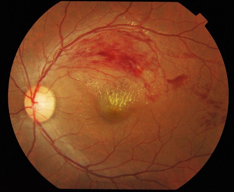
Retinal photo showing bleeding along an upper branch vein, with yellow deposits near the macula.
a pattern of retinal hemorrhage known as a 'flame shape appearance'
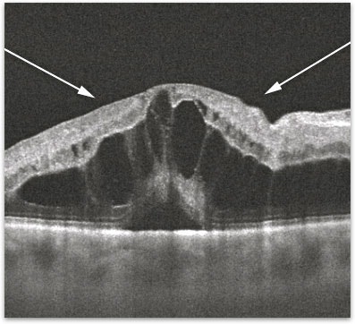
OCT scan of the retina; arrows point to the swollen retina, which contains dark fluid-filled spaces.
The OCT shows macular edema
This is a classic presentation of branch retinal vein occlusion (BRVO)
As the name suggests, a retinal vein has become blocked, causing bleeding
Our blood vessels are divided into arteries and veins.
Arteries are the vessels that supply blood to the retina,
and veins are the vessels that carry used blood from the retina back to the heart
To put it simply, arteries are like the water supply, and veins are like the drainage system.
When a branch retinal vein is blocked, it's like a clogged drain: blood can't flow down to the heart, so it backs up and gushes out, right?
This is a simplified analogy, but this is the mechanism that causes retinal hemorrhage.
Now, you might wonder: what happens when an artery is blocked?
In that case, it's called retinal artery occlusion, and it is an ophthalmic emergency.
Arteries are not easily blocked, so this condition is mainly caused by mechanical factors, such as filler injection procedures.
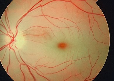
Fundus photo of the retina showing the optic disc on the left, branching blood vessels and the macula in the center.
Arteries supply clean blood and nutrients to the retinal tissue,
so when they are blocked, the retinal tissue that no longer receives blood and nutrients falls into ischemia (a state in which blood is not adequately supplied to the tissue).
In the image above, the 'cherry red spot' gets its name because the ischemic retina turns relatively white, making the central macula look like a red dot.
Retinal artery occlusion is an ophthalmic emergency, and various measures are taken to lower intraocular pressure, including IOP-lowering eye drops, ocular massage, anterior chamber paracentesis, and vitrectomy.
However, once an artery is blocked, reperfusion is rare, and unfortunately, the visual prognosis is not very good.
Now, back to branch retinal vein occlusion.
Let's look at why veins get blocked in the first place.
The most common cause of branch retinal vein occlusion is hypertension. Other causes, including arteriosclerosis, diabetes, and blood disorders, can also lead to retinal vascular occlusion.
In branch retinal vein occlusion, neovascularization may develop in the retinal area where bleeding occurred, as a result of ischemic changes.
To suppress this, focal laser photocoagulation may also be performed.
Intravitreal anti-VEGF antibody injections are used to suppress neovascularization and reduce macular edema.
Laser photocoagulation and intravitreal injections! If you've been diligently reading Seongmo Jin Eye Clinic's posts, you probably have a good idea by now, right?
That's right: these are the same injections used to suppress neovascularization in macular degeneration and diabetic macular edema.
Besides antibody injections, steroid injections can also be used to reduce macular edema.
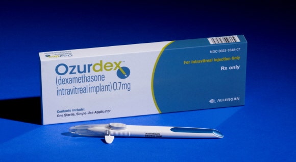
Ozurdex (dexamethasone intravitreal implant 0.7 mg) by Allergan: product box and single-use applicator.
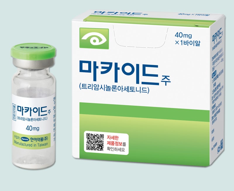
Maqaid Inj. (triamcinolone acetonide) 40 mg vial and box from Hanmi; a QR code links to detailed product information.
Steroids work well to reduce macular edema by acting on various cytokines, but because they can cause cataracts and raise intraocular pressure, they should be used appropriately after consultation with a retina specialist.
Unlike arterial occlusion, branch retinal vein occlusion can be treated effectively if you receive appropriate treatment at the right time.
It is possible to restore vision to a normal range by reducing macular edema and suppressing neovascularization.
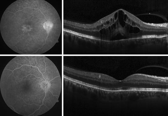
Angiography and OCT images: the top row shows macular swelling with fluid cysts; the bottom row shows a flatter retina without cysts.
Being treated by a skilled doctor is what matters most!
For more detailed information, please refer to the Seongmo Jin Eye Clinic website.
Presented with sincerity and skill, this has been Park Jin-hyeong, Chief Director of Seongmo Jin Eye Clinic ^^
#SeongmoJinEyeClinic #JinEyeClinic #RetinalVeinOcclusion #BranchRetinalVeinOcclusion #BRVO #Floaters #SeongmoJinEyeClinic #JinEyeClinic #MacularDegeneration #RetinalDisease #TrueEyeClinic #Retina #Ophthalmology #Presbyopia #IPL #SkinCare #TotalCare #Laser #AntiAging #AntiAging #Whitening #Cataract #MultifocalLens #CataractSurgery #GangnamEyeClinic #SeongmoEyeClinic #PresbyopiaSurgery #OphthalmologyProfessor #Glaucoma #DryEye #DryEyeSyndrome #DryEye #LASIK #LASEK #GangnamLASIK #SmileLASIK #ArtificialTears #Lens #PainFreeLASEK #TodayLASEK
Frequently Asked Questions
What is branch retinal vein occlusion?
Branch retinal vein occlusion is a condition in which a retinal vein becomes blocked, causing bleeding. Veins carry used blood from the retina back to the heart, and according to the article, when this pathway is blocked, blood flows back and causes retinal hemorrhage.
What are the main causes of branch retinal vein occlusion?
The most common cause is hypertension. Retinal vascular occlusion can also be caused by arteriosclerosis, diabetes, and blood disorders.
What symptoms does branch retinal vein occlusion cause?
In the case described in the article, the patient came in with sudden blurring in one eye. Testing showed reduced corrected visual acuity, fundus examination revealed flame-shaped retinal hemorrhage, and OCT showed macular edema.
How is branch retinal vein occlusion treated?
Treatment may include laser photocoagulation, intravitreal anti-VEGF injections, and steroid injections. Lasers and antibody injections are used to suppress neovascularization, and antibody injections are also used to reduce macular edema. Steroids reduce macular edema but can cause cataracts and raise intraocular pressure, so they should be used appropriately after consultation with a retina specialist.
Can vision recover with branch retinal vein occlusion?
Yes. With timely and appropriate treatment, it is possible to restore vision to a normal range. Unlike arterial occlusion, venous occlusion can improve with treatments that reduce macular edema and suppress neovascularization, and the article emphasizes that the timing of treatment matters.
How does retinal artery occlusion differ from vein occlusion?
According to the article, retinal artery occlusion is an ophthalmic emergency with a poor visual prognosis. In contrast, with branch retinal vein occlusion, vision can be restored to a normal range with timely treatment. Because arteries are not easily blocked, artery occlusion mainly occurs from mechanical causes such as filler injection procedures.